The Cuckoo Optimization Algorithm Enhanced Visualization of Morphological Features of Diabetic Retinopathy
DOI:
https://doi.org/10.37385/jaets.v4i2.1978Keywords:
Diabetic retinopathy, Fundus image, Cuckoo algoritm, Image EnhancementAbstract
This research compares strategies for identifying diabetic retinopathy (DR) using fundus image and discusses the efficiency of various image pre-processing techniques to enhance the quality of fundus images. Fundus images in medical image processing often suffer from non-uniform lighting, low contrast, and noise issues, which necessitate image pre-processing to enhance their quality. The study evaluates the effectiveness of several optimization techniques in selecting the best technique for identifying DR. One of the image pre-processing techniques compared in the study involves comparing negative images, dark contrast stretch, light contrast stretch, and partial contrast stretch, which are then evaluated using standard performance metrics such as NIQE, PNSR, MSE, and entropy. The results are further optimized using the Cuckoo Search Algorithm. The proposed technique produces better image quality improvements in several performance metrics, such as MSE, NIQE, PSNR, and entropy. Bright Contrast Stretch outperforms other techniques in NIQE Mean 5.2850, Entropy 5.0193, NIQE Standard deviation 0.2261, and Entropy 0.2612.
Downloads
References
Abdul-nasir, A. S., Mashor, M. Y., & Mohamed, Z. (2013). Colour Image Segmentation Approach for Detection of Malaria Parasites Using Various Colour Models and k -Means Clustering. 10(1), 41–55.
Acharya, U. K., & Kumar, S. (2021). Genetic algorithm based adaptive histogram equalization (GAAHE) technique for medical image enhancement. Optik, 230(January), 166273. https://doi.org/10.1016/j.ijleo.2021.166273
Alyoubi, W. L., Shalash, W. M., & Abulkhair, M. F. (2020). Diabetic retinopathy detection through deep learning techniques: A review. Informatics in Medicine Unlocked, 20(03). https://doi.org/10.1016/j.imu.2020.100377
Astorga, J. E. O., Wang, L., Yamada, S., Fujiwara, Y., Du, W., & Peng, Y. (2022). Automatic Detection of Microaneurysms in Fundus Images. International Journal of Software Innovation, 11(1), 1–14. https://doi.org/10.4018/IJSI.315658
Bennet, M. A., Dharini, D., Priyadharshini, S. M., & Mounica, N. L. (2016). Detection of blood vessel Segmentation in retinal images using Adaptive filters. 8(4), 290–298.
Bhateja, V., Satapathy, S. C., Travieso-González, C. M., & Aradhya, V. N. M. (2021). Correction to: Data Engineering and Intelligent Computing. https://doi.org/10.1007/978-981-16-0171-2_61
Bhimavarapu, U., & Battineni, G. (2022). Automatic Microaneurysms Detection for Early Diagnosis of Diabetic Retinopathy Using Improved Discrete Particle Swarm Optimization. Journal of Personalized Medicine, 12(2). https://doi.org/10.3390/jpm12020317
Chen, L. (2022). Application of Cuckoo Search Algorithm in Cost Estimation of Building Energy Engineering. Wireless Communications and Mobile Computing, 2022. https://doi.org/10.1155/2022/7956751
Datta, N. S., Dutta, H. S., De, M., & Mondal, S. (2013). An Effective Approach: Image Quality Enhancement for Microaneurysms Detection of Non-dilated Retinal Fundus Image. Procedia Technology, 10, 731–737. https://doi.org/10.1016/j.protcy.2013.12.416
Deng, J., Tang, P., Zhao, X., Pu, T., Qu, C., & Peng, Z. (2022). Local Structure Awareness-Based Retinal Microaneurysm Detection with Multi-Feature Combination. Biomedicines, 10(1), 1–15. https://doi.org/10.3390/biomedicines10010124
Firdausy, K., Sutikno, T., & Prasetyo, E. (2007). Image Enhancement Using Contrast Stretching on Rgb and Ihs Digital Image. TELKOMNIKA (Telecommunication Computing Electronics and Control), 5(1), 45. https://doi.org/10.12928/telkomnika.v5i1.1335
Gayathri, K., Narmadha, D., Thilagavathi, K., Pavithra, K., & Pradeepa, M. (2014). Detection of Dark Lesions from Coloured Retinal Image Using Curvelet Transform and Morphological Operation. 2, 15–21.
Jadhav, A. S., & Patil, P. B. (2018). Detection of blood vessels in retinal images for diagnosis of diabetics. Proceedings of the 2nd International Conference on Inventive Systems and Control, ICISC 2018, Icisc, 888–891. https://doi.org/10.1109/ICISC.2018.8398928
Kaur, I., & Singh, L. M. (2016). A Method of Disease Detection and Segmentation of Retinal Blood Vessels using Fuzzy C-Means and Neutrosophic Approach. Imperial Journal of Interdisciplinary Research (IJIR), 2(6), 551–557.
Kusuma Whardana, A., & Suciati, N. (2014). A Simple Method for Optic Disk Segmentation from Retinal Fundus Image. International Journal of Image, Graphics and Signal Processing, 6(11), 36–42. https://doi.org/10.5815/ijigsp.2014.11.05
Maheswari, M. S., & Punnolil, A. (2014). A novel approach for retinal lesion detection in diabetic retinopathy images. People, 4(6).
Maini, R., & Aggarwal, H. (2010). A Comprehensive Review Of Image Enhancement Techniques. International Journal of Innovative Research and Growth, 8(6), 8–13. https://doi.org/10.26671/ijirg.2019.6.8.101
Mayya, V., Kamath S?, S., & Kulkarni, U. (2021). Automated microaneurysms detection for early diagnosis of diabetic retinopathy: A Comprehensive review. Computer Methods and Programs in Biomedicine Update, 1, 100013. https://doi.org/10.1016/j.cmpbup.2021.100013
Mazlan, N., Yazid, H., & Sabri, N. R. (2018). Enhancement of Retinal Images for Microaneurysms Detection in Diabetic Retinopathy. 2018 IEEE 16th Student Conference on Research and Development, SCOReD 2018, 1–5. https://doi.org/10.1109/SCORED.2018.8711081
Munteanu, C., & Rosa, A. (2000). Towards automatic image enhancement using Genetic Algorithms. Proceedings of the IEEE Conference on Evolutionary Computation, ICEC, 2, 1535–1542. https://doi.org/10.1109/cec.2000.870836
Pendekal, M. J., & Gupta, S. (2022). An Ensemble Classifier Based on Individual Features for Detecting Microaneurysms in Diabetic Retinopathy. Indonesian Journal of Electrical Engineering and Informatics, 10(1), 60–71. https://doi.org/10.52549/ijeei.v10i1.3522
Pundikal, M., & Holi, M. S. (2022). Microaneurysms Detection Using Grey Wolf Optimizer and Modified K-Nearest Neighbor for Early Diagnosis of Diabetic Retinopathy. International Journal of Intelligent Engineering and Systems, 15(1), 130–140. https://doi.org/10.22266/IJIES2022.0228.13
Ramasubramanian, B., & Selvaperumal, S. (2016). A comprehensive review on various preprocessing methods in detecting diabetic retinopathy. International Conference on Communication and Signal Processing, ICCSP 2016, 642–646. https://doi.org/10.1109/ICCSP.2016.7754220
Rosas-Romero, R., Martínez-Carballido, J., Hernández-Capistrán, J., & Uribe-Valencia, L. J. (2015). A method to assist in the diagnosis of early diabetic retinopathy: Image processing applied to detection of microaneurysms in fundus images. Computerized Medical Imaging and Graphics, 44, 41–53. https://doi.org/10.1016/j.compmedimag.2015.07.001
Salazar-Gonzalez, A., Kaba, D., Li, Y., & Liu, X. (2014). Segmentation of the blood vessels and optic disk in retinal images. IEEE Journal of Biomedical and Health Informatics, 18(6), 1874–1886. https://doi.org/10.1109/JBHI.2014.2302749
Salihah, A. N. A., Mashor, M. Y., Harun, N. H., & Rosline, H. (2010). Colour Image Enhancement Techniques for Acute Leukaemia Blood Cell Morphological Features. 3677–3682.
Sengupta, S., Singh, A., Leopold, H. A., Gulati, T., & Lakshminarayanan, V. (2020). Ophthalmic diagnosis using deep learning with fundus images – A critical review. In Artificial Intelligence in Medicine. https://doi.org/10.1016/j.artmed.2019.101758
Shiralkar, S., Bahulekar, A., & Jawade, S. (2022). The Cuckoo Search Algorithm: A review. International Research Journal of Engineering and Technology, 1238–1246. www.irjet.net
Soares, I., Castelo-Branco, M., & Pinheiro, A. (2023). Microaneurysms detection in retinal images using a multi-scale approach. Biomedical Signal Processing and Control, 79(P2), 104184. https://doi.org/10.1016/j.bspc.2022.104184
Subramanian, S., Mishra, S., Patil, S., Shaw, K., & Aghajari, E. (2022). Machine Learning Styles for Diabetic Retinopathy Detection: A Review and Bibliometric Analysis. Big Data and Cognitive Computing, 6(4). https://doi.org/10.3390/bdcc6040154
Sun, Y., Zhao, Z., Jiang, D., Tong, X., Tao, B., Jiang, G., Kong, J., Yun, J., Liu, Y., Liu, X., Zhao, G., & Fang, Z. (2022). Low-Illumination Image Enhancement Algorithm Based on Improved Multi-Scale Retinex and ABC Algorithm Optimization. Frontiers in Bioengineering and Biotechnology, 10(April), 1–16. https://doi.org/10.3389/fbioe.2022.865820
Swathi, C., Anoop, B. K., Dhas, D. A. S., & Sanker, S. P. (2017). Comparison of different image preprocessing methods used for retinal fundus images. 2017 Conference on Emerging Devices and Smart Systems, ICEDSS 2017, October 2017, 175–179. https://doi.org/10.1109/ICEDSS.2017.8073677
Tavakoli, M., Mehdizadeh, A., Aghayan, A., Shahri, R. P., Ellis, T., & Dehmeshki, J. (2021). Automated Microaneurysms Detection in Retinal Images Using Radon Transform and Supervised Learning: Application to Mass Screening of Diabetic Retinopathy. IEEE Access, 9, 67302–67314. https://doi.org/10.1109/ACCESS.2021.3074458
Toresa, D., Azrul, M., Shahril, E., Hazlyna, N., Abu, J., & Amnur, H. (2021). Automated Detection and Counting of Hard Exudates for Diabetic Retinopathy by using Watershed and Double Top-Bottom Hat Filtering Algorithm. 5(September), 242–247.
Yadav, D., Karn, A. K., Giddalur, A., Dhiman, A., Sharma, S., Muskan, & Yadav, A. K. (2021). Microaneurysm detection using color locus detection method. Measurement: Journal of the International Measurement Confederation, 176(July 2020), 109084. https://doi.org/10.1016/j.measurement.2021.109084
Zhang, M., Jiang, W., Zhou, X., Xue, Y., & Chen, S. (2019). A hybrid biogeography-based optimization and fuzzy C-means algorithm for image segmentation. Soft Computing, 23(6), 2033–2046. https://doi.org/10.1007/s00500-017-2916-9

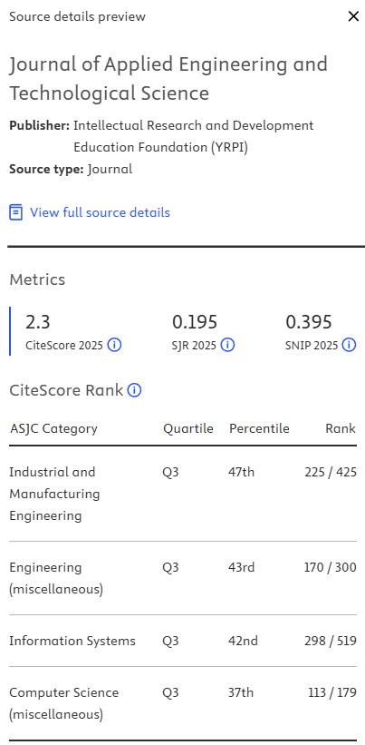


 CITEDNESS IN SCOPUS
 CITEDNESS IN WOS


